Muscle Breakdown Biceps Femoris
Biceps femoris Origin, insertion, innervation, function Kenhub
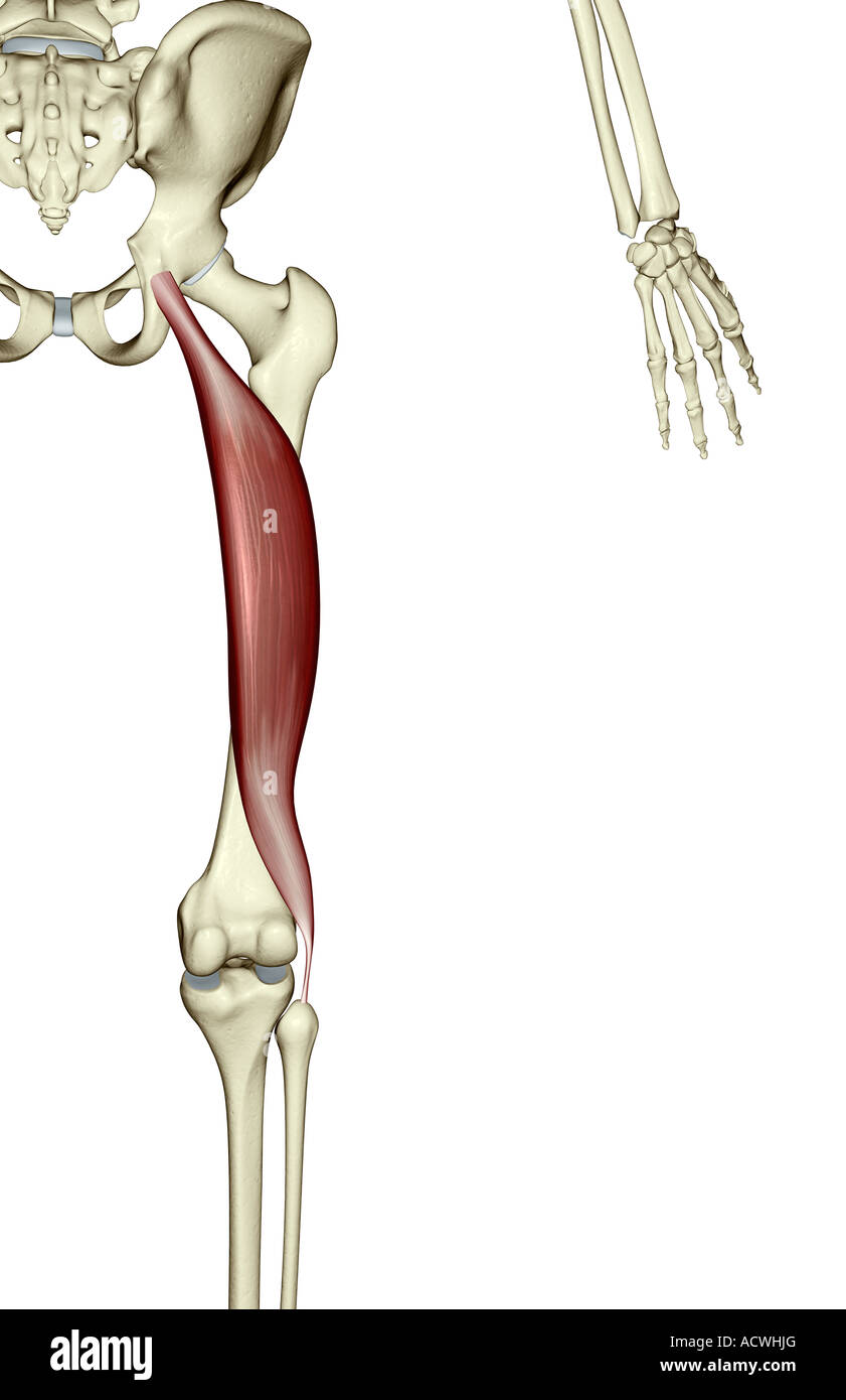
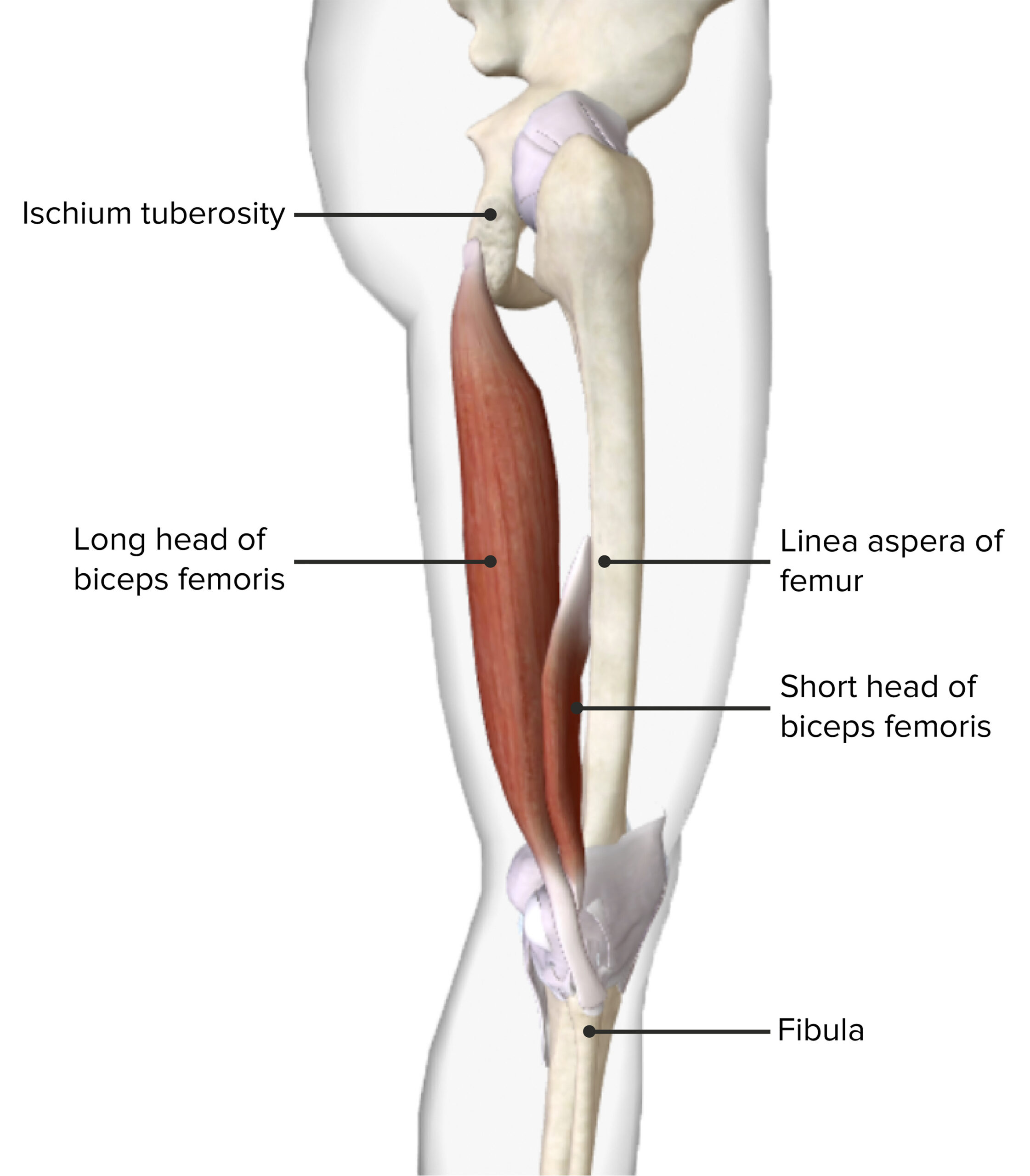
The biceps femoris tendon attaches the muscle bellies of the long and short heads of the biceps femoris muscle to the head of the fibula. This insertion site allows the biceps femoris muscle to be able to: - flex the leg at the knee joint; - laterally rotate the leg at the knee joint while this joint is held in a semiflexed position.
Biceps Femoris Rehab My Patient
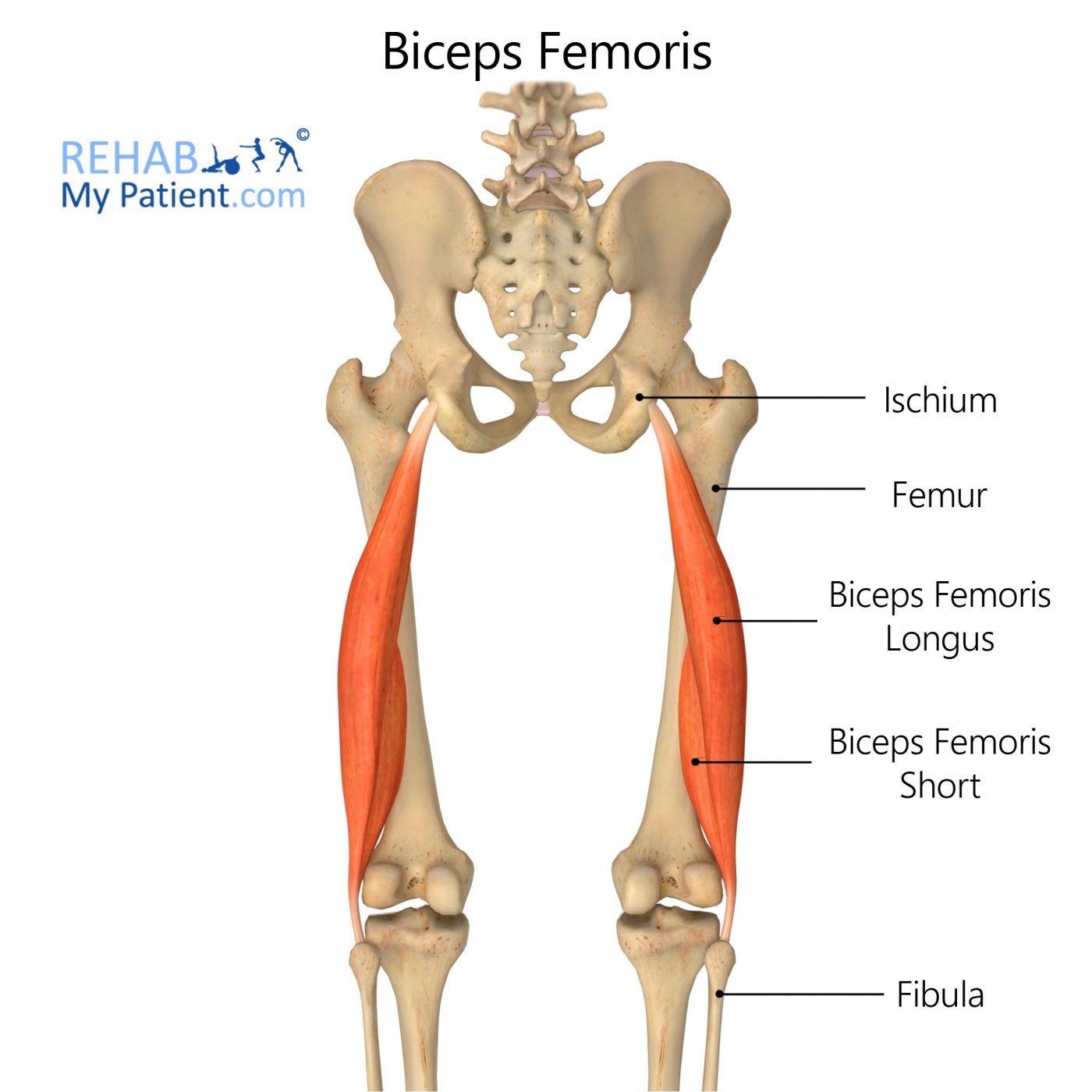
The biceps femoris is a double-headed muscle located on the back of thigh. It consists of two parts: the long head, attached to the ischium (the lower and back part of the hip bone), and the.
Biceps Femoris Longus Muscle Photograph by Sebastian Kaulitzki/science Photo Library Fine Art
M. biceps femoris: The biceps femoris muscle arises from the ischial tuberosity. The muscle forms a thin aponeurosis that is inserted into the deep fascia of the proximal hind limb, the head of the fibula, the lateral tibial condyle, and the capsule of the knee joint. This muscle can be used to administer intramuscular injections [12]. •

Základy sportovní kineziologie Fakulta sportovních studií
Dr. Ebraheim's educational animated video describes the condition of biceps femoris muscle anatomy.Follow me on twitter:https://twitter.com/#!/DrEbraheim_UTM.

Biceps Femoris Anatomy Origin, Insertion & Action YouTube
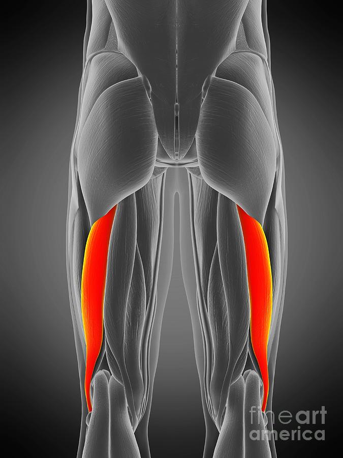
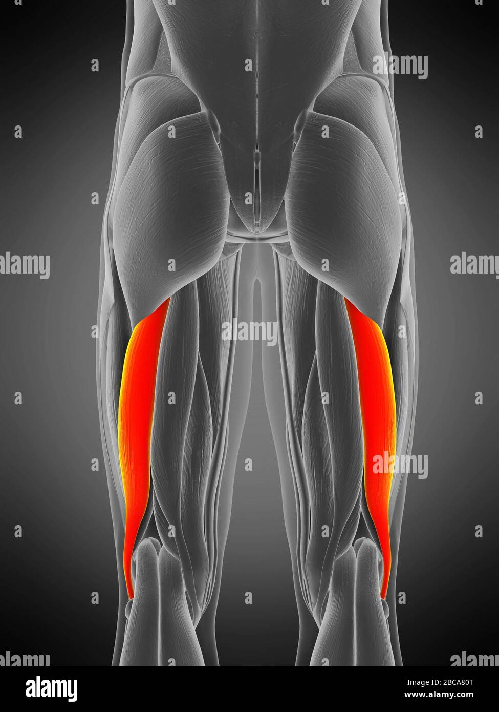
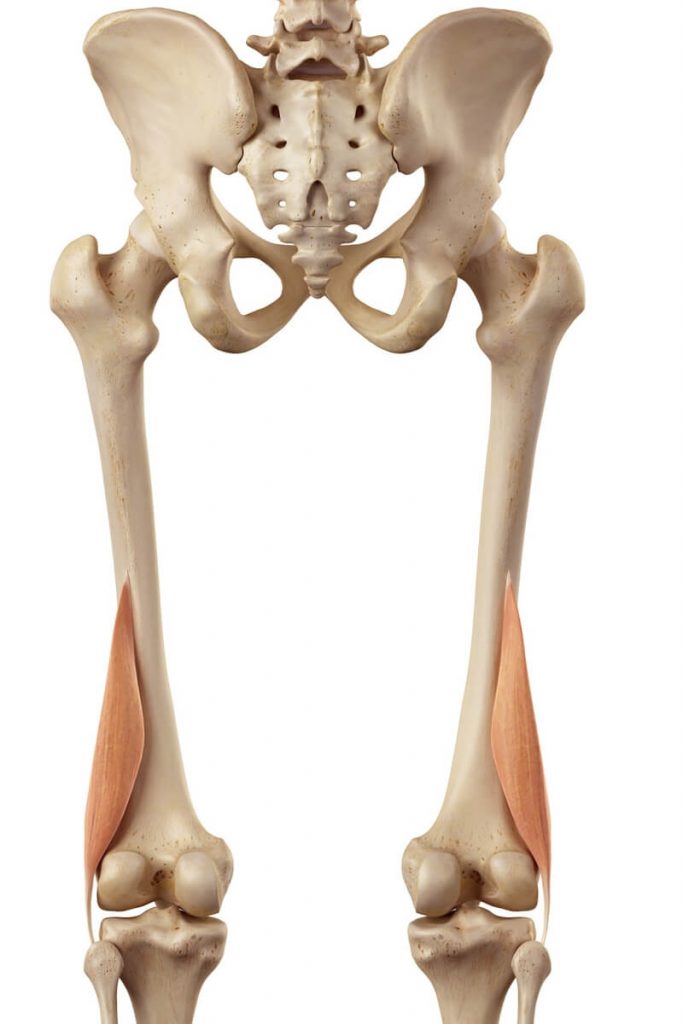
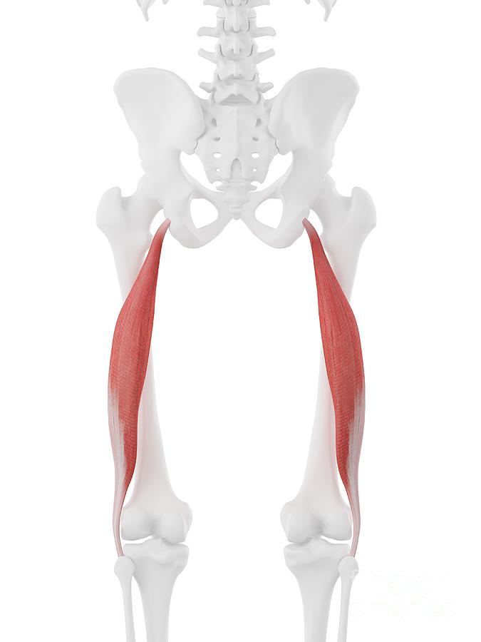
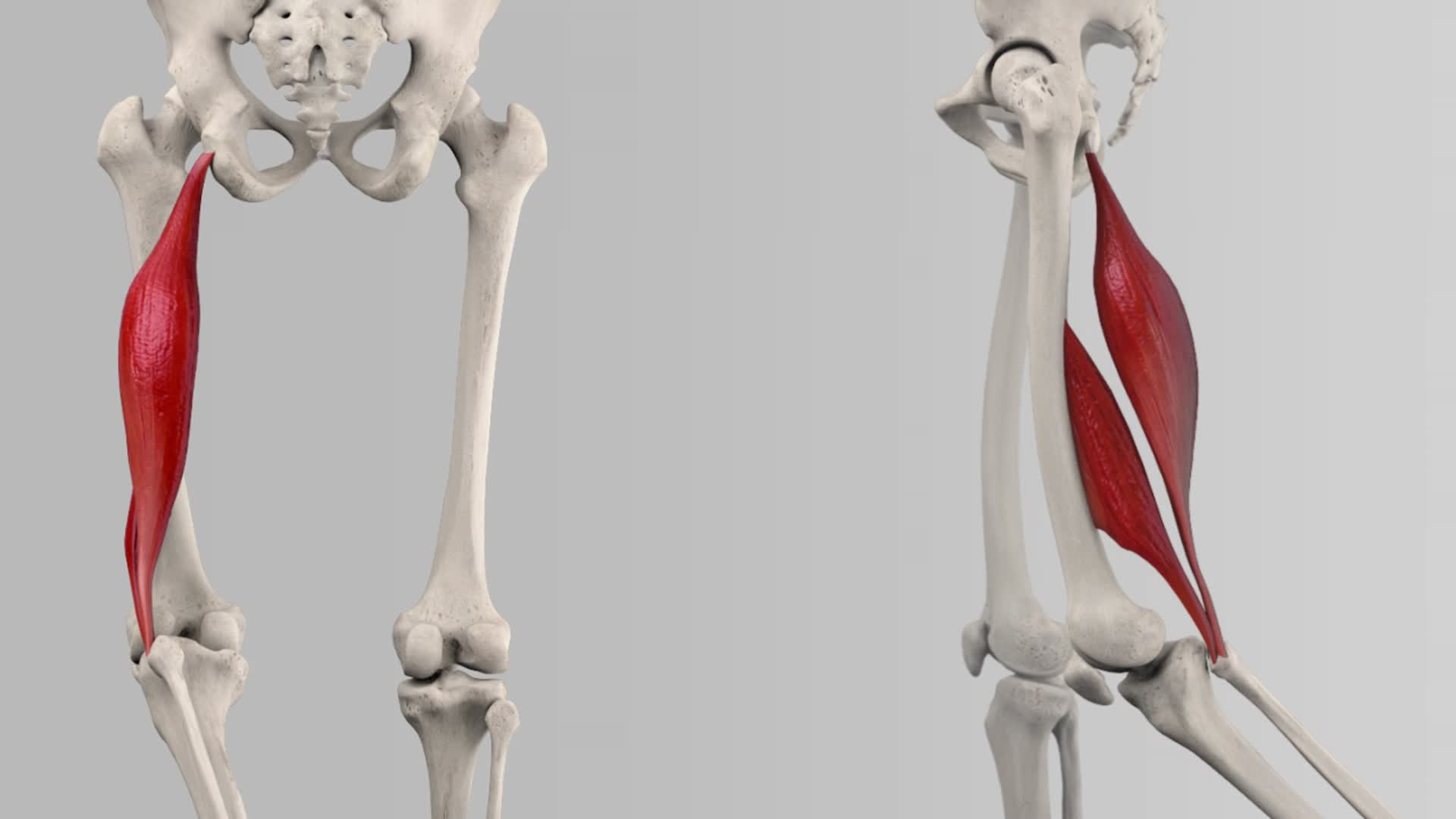
Biceps femoris is a long muscle of the posterior aspect of the thigh. Together with the semitendinosus and semimembranosus muscles, it makes the group of muscles commonly known as the hamstrings. The biceps femoris muscle runs from the ischial tuberosity, all the way to the proximal part of the fibula.
Biceps femoris fotografías e imágenes de alta resolución Alamy
Dr. Ebraheim's educational animated video describes the condition of biceps femoris muscle anatomy. The biceps femoris is a muscle of the posterior thigh. The biceps femoris is one of the three flexor muscles of the posterior thigh. The ischial tuberosity is divided by a transverse ridge into an upper quadrangular portion and a lower.
Músculo bíceps femoris largo, ilustración Fotografía de stock Alamy
The simple fibular and tibial repair construct () involved 1 double-loaded 4.5-mm suture anchor (Corkscrew) at the proximal fibular insertion of the biceps femoris, one 3.0-mm suture anchor (SutureTak) at the distal fibular insertion of the biceps femoris, and one 3.0-mm suture anchor (SutureTak) at the tibial insertion of the biceps femoris.

Anatomical model showing the biceps femoris muscles Stock Photo Alamy
Avulsion injury of the distal biceps femoris can occur as an isolated injury or as part of a multiligament injury pattern, with or without concomitant injury to additional lateral knee structures. ∥ Through qualitative, quantitative, and biomechanical study, the anatomy and strength of the fibular collateral ligament (FCL), popliteofibular ligament, and popliteus tendon have been described.

biceps origin and insertion
The biceps femoris is a long muscle in the posterior compartment of the thigh responsible for movement at both the hip and knee joints. Along with the semitendinosus and semimembranosus, the biceps femoris makes up the hamstrings muscle. The muscles of the hamstring border the popliteal fossa, which is a triangular space behind the knee.

Muscle Breakdown Biceps Femoris
In more technical terms, the biceps femoris is a two-headed (hence "biceps") skeletal muscle, with one superficial head and the other being far deeper in the leg. The origin point is in the femur, while the distal attachment point lies in the fibula of the calves. The biceps femoris is located alongside the semitendinosus and.
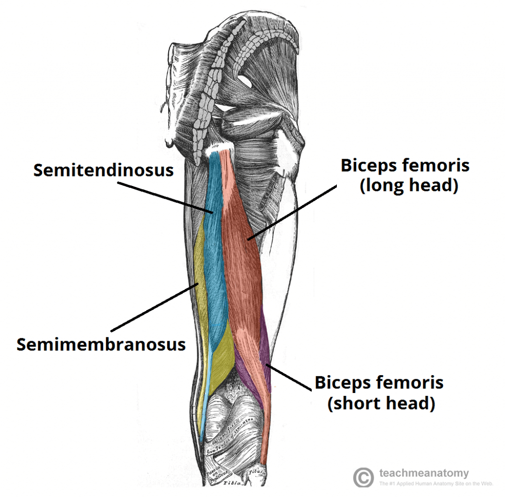
Biceps Femoris Attachments Actions TeachMeAnatomy
Definition The biceps femoris is a muscle found at the back of the thigh. Part of the biceps femoris belongs to the hamstrings muscle group. The biceps femoris is a two-part spindle-shaped muscle. It has two heads: the long head and the short head.

Biceps femoris What Is It, Location, Action, and More Osmosis
The biceps femoris muscle contributes to the formation of the popliteal fossa, where the muscle and tendon form its superolateral boundary. The term "hamstrings" is the collective name given to the long head of biceps femoris, semitendinosus, and semimembranosus muscles. These three muscles share similar features that the short head of.
Musculus biceps femoris sportbachelor
M. biceps femoris: The biceps femoris muscle arises from the ischial tuberosity. The muscle forms a thin aponeurosis that is inserted into the deep fascia of the proximal hind limb, the head of the fibula, the lateral tibial condyle, and the capsule of the knee joint.
Biceps Femoris Longus Muscle Photograph by Sebastian Kaulitzki/science Photo Library Pixels
Dr. Ebraheim's educational animated video describes the anatomy of the Biceps Femoris muscle. The biceps femoris is a muscle of the posterior thigh. The bic.
Thigh Anatomy Concise Medical Knowledge
The biceps femoris muscle is the strongest of the hamstring complex and is responsible for flexion, external rotation, and posterolateral stability of the knee. The biceps femoris muscle has a long and a short head. In some normal variants, the short head of the biceps femoris may be absent.
Musculus biceps femoris DocCheck
The biceps femoris is one of the large muscles in the posterior compartment of the thigh and a component of the hamstrings. It has a long and a short head, each with different functions and innervation. Its medial border forms the superolateral border of the popliteal fossa. Summary origin long head: medial facet of the ischial tuberosity
